Ultrasound is one of the most commonly used tests in medicine. Indeed, it allows the diagnosis of many disorders such as malformations, tumors or cysts. It is also crucial to visually guide the physician during the biopsy procedure or other examinations. Let's take a look at this method of medical examination.
The different categories of an ultrasound
Medical ultrasound is divided into two distinct categories: diagnostic and therapeutic.
Diagnostic ultrasound
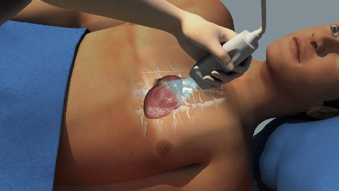
Diagnostic ultrasound is a noninvasive diagnostic technique used to produce images in the body. Ultrasound probes, called transducers, produce sound waves with frequencies above the threshold of the human ear. Most diagnostic ultrasound probes are placed on the skin.
However, to optimize image quality, probes can be deposited inside the body through the gastrointestinal tract, vagina or blood vessels. In addition, ultrasound is sometimes used during surgery by placing a sterile probe in the area where the operation is being performed.
It can also be subdivided into anatomical and functional ultrasound. Anatomical ultrasound produces images of internal organs or other structures. Functional ultrasound combines information such as tissue or blood movement and velocity, tissue softness or hardness, and other physical characteristics with anatomical images to create "information maps." These maps help physicians visualize changes/differences in function within a structure or organ.
Therapeutic ultrasound
Therapeutic ultrasound also uses sound waves superior to the human ear, but does not produce images. Its purpose is to interact with the body's tissues so that they can be modified or destroyed. Possible modifications include: moving or pushing tissue, heating it, dissolving clots, or administering medications to specific sites in the body.
These destructive or ablative functions are possible by using very high intensity rays that can destroy diseased or abnormal tissue such as tumors. The use of ultrasound has the advantage of not being invasive in most cases. There is no need to make cuts or incisions in the skin, so no wounds or scars remain.
How is the ultrasound scan performed?
Ultrasound waves are produced by a transducer that can emit ultrasound waves and detect the echoes reflected by the ultrasound.
The active elements of the ultrasonic transducers are made of special glass ceramic materials called piezoelectric. These materials are capable of producing sound waves when an electric field passes through them. But they also work in the opposite direction, producing an electric field when they receive a sound wave.
When used in an ultrasound scanner, the transducer sends a beam of sound waves into the body. The sound waves are reflected back to the transducer through the tissue boundaries in the beam path. When these echoes reach the transducer, electrical signals are generated and sent to the ultrasound machine.
An ultrasonic transducer
Using the speed of sound and the return time of each echo, the scanner calculates the distance between the transducer and the tissue boundary. These distances are then used to generate two-dimensional images of tissues and organs. During an ultrasound, the technician will apply a gel to the skin. This prevents air pockets from forming between the transducer and the skin. This can prevent the ultrasound from penetrating the body.
What is the purpose of ultrasound?
Diagnostic ultrasound
Diagnostic ultrasound is capable of producing images of the body's internal organs in a non-invasive manner. However, it is not good for imaging bone or air-containing tissue, such as lungs. Under certain conditions, ultrasound can image bones or lungs and the membrane that covers them when they are full or partially filled with fluid.
One of the most common uses of ultrasound is during pregnancy to monitor fetal growth and development. However, it has many other uses, including the production of images of the heart, blood vessels, eyes, thyroid, brain and thorax... Ultrasound images are displayed in 2D, 3D or 4D (3D in motion).
Functional ultrasound
Applications of functional ultrasound include color Doppler ultrasound to measure and visualize blood flow in body vessels or the heart. It can also measure the speed of blood flow and the direction of movement. This is done using color-coded maps called color Doppler images. Doppler ultrasonography is commonly used to determine whether plaque accumulation in the carotid arteries blocks the blood flow to the brain.
Ultrasound is also an important method for producing images of interventions in the body. For example, the biopsy Ultrasound-guided needle aspiration helps physicians determine the position of a needle while being guided to a selected target, such as a breast mass or tumor.
Similarly, ultrasound can provide real-time images of the position of a catheter tip as it is inserted into a blood vessel and guided along the vessel. It can also be used in minimally invasive surgery to guide the surgeon with images of the inside of the body in real time.
Therapeutic or interventional ultrasound
Therapeutic ultrasound produces high levels of acoustic response. They can be focused on specific objectives regarding the effects of heating, ablation or rupture of a tissue. One type of therapeutic ultrasound uses highly focused, high-intensity sound beams called high-intensity focused ultrasound.
This type of ultrasound is currently FDA approved for the treatment of uterine fibroids, for pain relief from bone metastases, and more recently for the removal of prostate tissue. It is also being studied to close wounds and stop bleeding, dissolve blood clots in blood vessels and temporarily open the blood-brain barrier so that drugs can enter.
Are there any risks?
Diagnostic ultrasound is generally considered safe and does not produce ionizing radiation such as that produced by X-rays. However, ultrasound can produce certain biological effects on the body under specific conditions and environments.
For this reason, the FDA requires that diagnostic ultrasound devices operate within acceptable limits. The FDA, as well as many professional societies, advises against the occasional use of ultrasound (e.g., for souvenir videos). She recommends using them only when there is a real medical need.